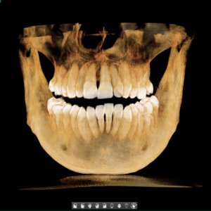
Panoramic Dental X-ray – When, Why, and How?
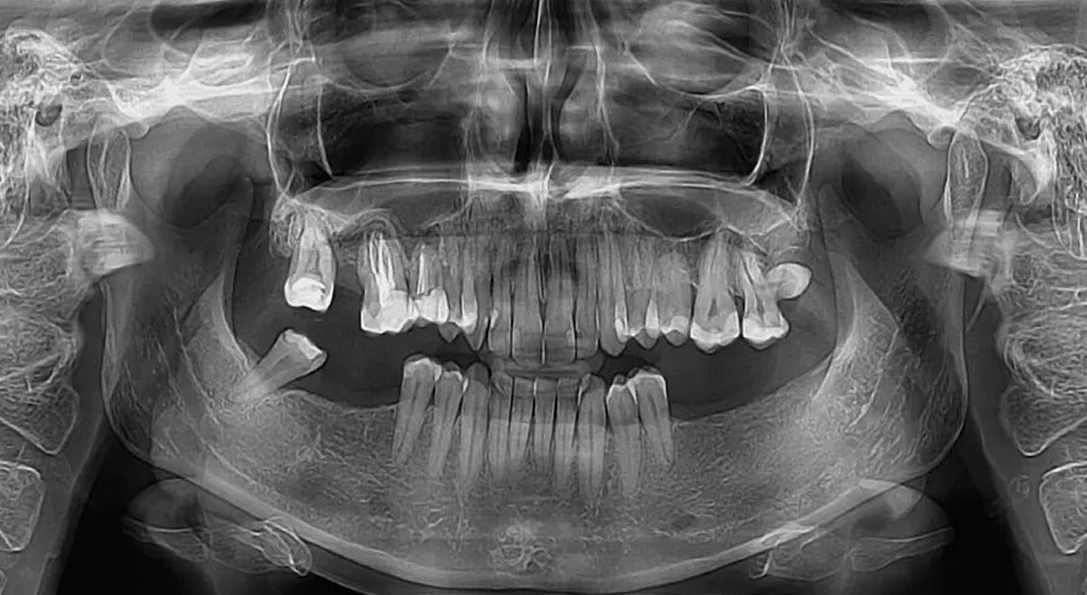
Next-Generation Panoramic Dental X-ray (OPG)
In modern dentistry, a visual examination with a mirror is only the tip of the iceberg. What happens beneath the gums, within the bone, and in the jaw joints often determines the success of the entire treatment. That is why panoramic dental imaging — Orthopantomography (OPG) — has become the gold standard and an essential tool for comprehensive treatment planning.
At our center in Belgrade, we have raised the bar to the highest level by investing in the latest-generation equipment, offering a level of image resolution previously unavailable on the local market.
What Is a Panoramic Dental X-ray (Orthopantomography)?
From a professional standpoint, an OPG is a panoramic radiographic image of the jaws taken in an orthoradial projection. The key characteristic of this method is that the central X-ray beam remains perpendicular to the plane of the jaws throughout the entire scanning process.
While the X-ray tube moves behind the patient’s head along a parabolic path, digital sensors simultaneously capture data in front of the head. The result is a comprehensive and accurate visualization of:
- The entire mandible with both temporomandibular joint condyles
- Maxillary sinuses (especially the alveolar recesses)
- The maxilla with all associated teeth
- Salivary gland regions
Technology That Makes the Difference: Dental Scan vs. Older Systems
As professionals, we are well aware that the main drawbacks of older panoramic machines were image distortion in the premolar region and the “ghosting” or overlapping of the cervical spine over the anterior teeth.
Our new equipment eliminates these limitations:
- High resolution without magnification: Older systems often caused magnification and loss of sharpness due to the distance between the cassette and the object. Our next-generation digital sensors provide true 1:1 proportions.
- Perfect tomographic focal layer: Our system uses three centers of rotation, allowing the sharp imaging zone (approximately 1 cm wide) to precisely follow the anatomical curvature of the patient’s jaw.
- Reduced artifacts: Thanks to advanced algorithms, oropharyngeal brightness and soft tissue shadows are minimized, resulting in clearer diagnostic images.
Indications: When Is a Panoramic X-ray Essential?
Whether you are a patient seeking a healthy smile or a dentist planning therapy, an OPG scan is indispensable for:
- Implantology and oral surgery – planning dental implant placement and impacted wisdom tooth surgery
- Pathological conditions – detection of cysts, tumors, and inflammatory bone lesions
- General dentistry – caries detection, restoration evaluation, and follow-up of endodontically treated teeth
- Orthodontics – monitoring tooth development in children and planning fixed orthodontic appliances
- Periodontology – precise assessment of alveolar bone levels around all teeth simultaneously
- TMJ diagnostics – comparative visualization of both temporomandibular joint condyles
What Patients Should Know
The imaging procedure itself is fast, safe, and comfortable.
- Preparation: Before the scan, all jewelry, earrings, glasses, and piercings must be removed, as metal objects can create “ghost images” that obscure important anatomical structures.
- Positioning: The head is stabilized at three points to ensure maximum precision.
- Duration: The scan takes 15 to 19 seconds, during which it is important to remain completely still.
Conclusion: Image Quality Directly Affects Treatment Quality
Not all panoramic X-rays are the same. A poor-quality image can conceal early root pathology or misrepresent bone density. By choosing our center, you are choosing the most advanced dental diagnostics in Belgrade. Our equipment is not only new — it represents the current technological peak in dental radiology.
Get your panoramic dental X-ray immediately — no appointment required!
Venizelosova 3, Stari Grad, Belgrade
For more information, call or send us a message.